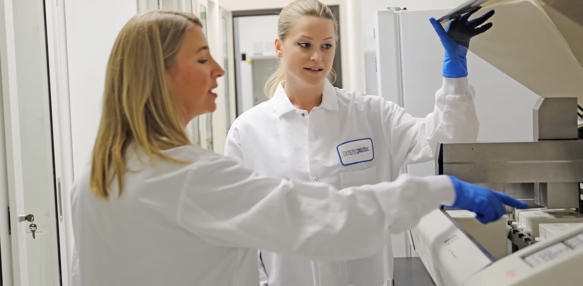
PathXdx technologists in lab coats and gloves collaborating at laboratory instrumentation

Research Pathology · Preclinical & Translational

More than histology. Quantitative answers from tissue.

PathXDx is a Brisbane, California research pathology laboratory supporting biotech, pharma, CRO, and academic programs, from routine histology and IHC through multiplex immunofluorescence, whole-slide imaging, and quantitative image analysis.

Owned scanning and in-house image analysis mean your slides don't leave our lab, and your data doesn't wait on a third party.

Scope
Research use only
Capabilities
Histology · IHC · Multiplex IF · Digital pathology
Imaging
Whole-slide scanning & analysis, in-house
Location
Brisbane, California
Model
Dedicated technologist per program
Reach
Mon–Fri 7am–7pm · Sat 9am–5pm · 24/7 contact

Why research teams work with us

High-touch partnership, owned imaging capability

  • Imaging and analysis under one roof

    We own our scanning and run image analysis in-house. That means no pass-through vendors, no queue behind someone else's work, and a single point of accountability from block to quantified result.

  • Multiplex without building it yourself

    Multiplex immunofluorescence takes real optimization to get right. We've done that work, so you don't need to stand up the capability internally for a single study.

  • A technologist who knows your protocol

    You get named technologists assigned to your program rather than whoever is free. They learn your tissue, your protocol, and your acceptance criteria.

  • Straight answers on scope

    If a study design won't produce the data you need, we say so before you commit, not after.

In the lab

Precision pathology starts with people who sweat the details

The same care that goes into your slides shows up in how we communicate: clear milestones, realistic timelines, and a single thread from study design through quantified delivery.

  • QA-minded handling from accessioning through cover-slipping
  • Multiplex IF, IHC, whole-slide imaging, and in-house analysis
  • Transparent proposals: PO and timeline, then execution
PathXdx laboratory professional in a white coat and safety glasses adjusting protective eyewear beside instrumentation

Capabilities

Differentiated services first, digital pathology and multiplex, then the full research pathology workflow.

  • Digital Pathology & Image Analysis

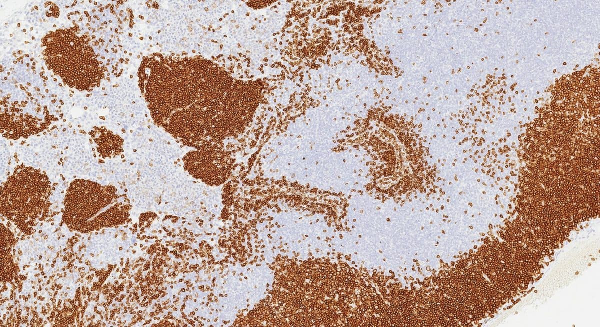
    Whole-slide scanning and quantitative image analysis, performed in-house on our own equipment. Brightfield and fluorescence scanning, digital slide management, and quantitative readouts: cell density, marker positivity, co-expression, and spatial relationships between populations. Because analysis is digital, we can support programs anywhere, including collaborators outside the US who send images rather than tissue.

  • Multiplex Immunohistochemistry & Immunofluorescence

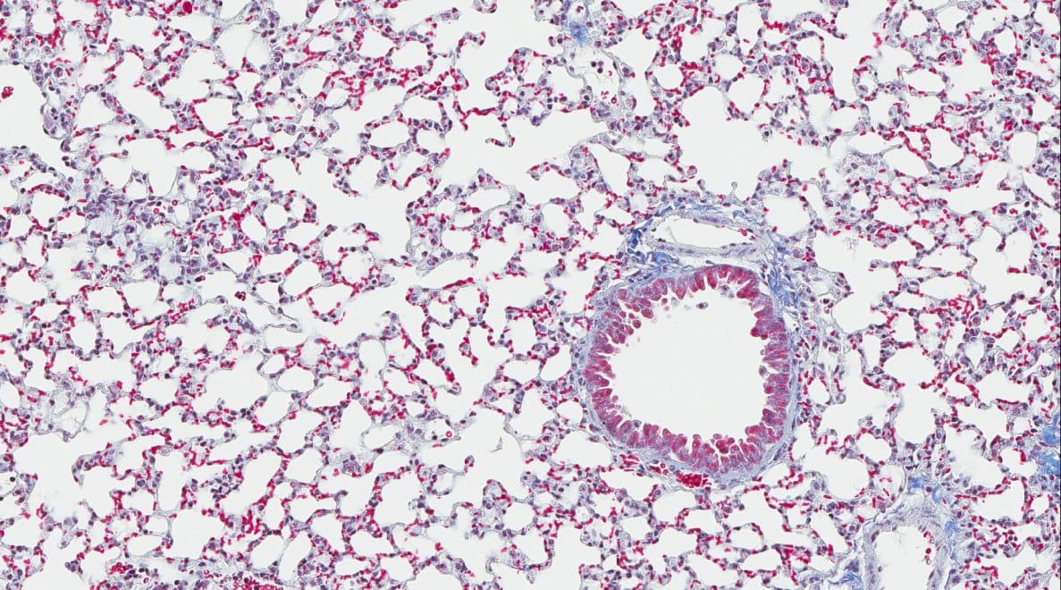
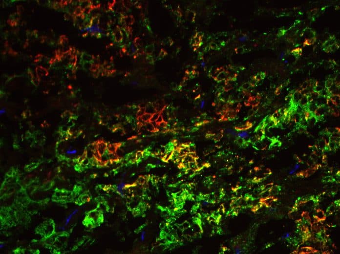
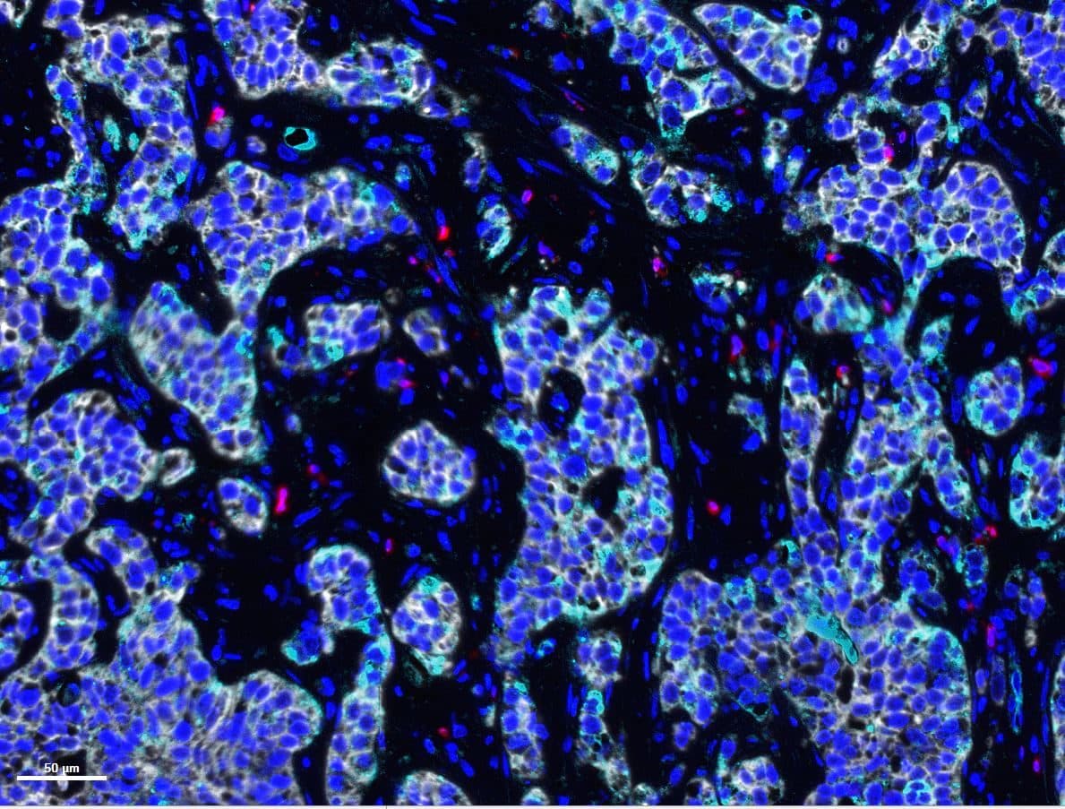
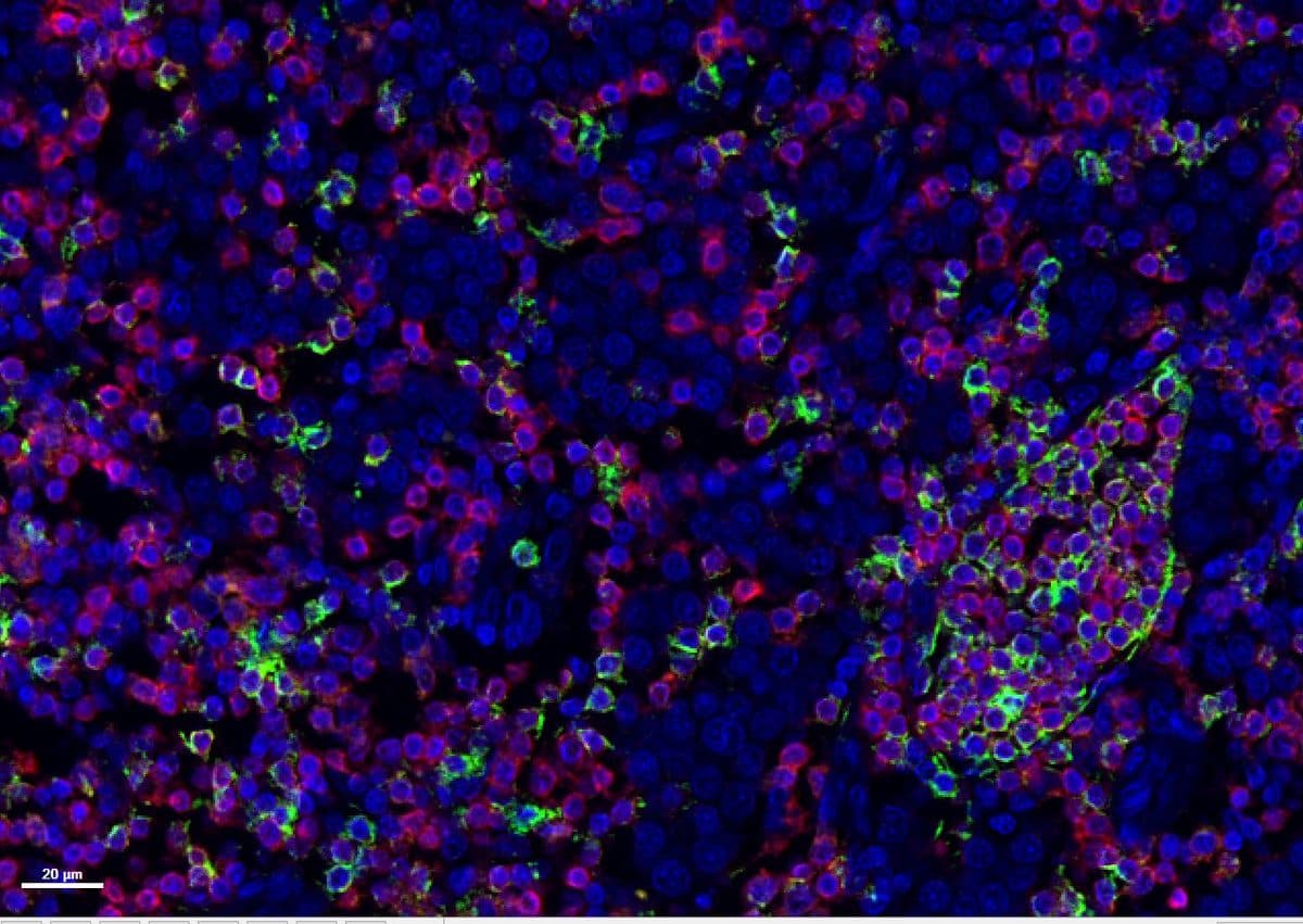
    Multiplex immunofluorescence panels at 3–4 plex, developed and optimized for your targets and tissue. Panel design, antibody optimization, staining, imaging, and quantitative analysis delivered as one workflow rather than handed between vendors. Useful when single-marker IHC can't answer the question: immune profiling, co-expression, and tissue microenvironment characterization.

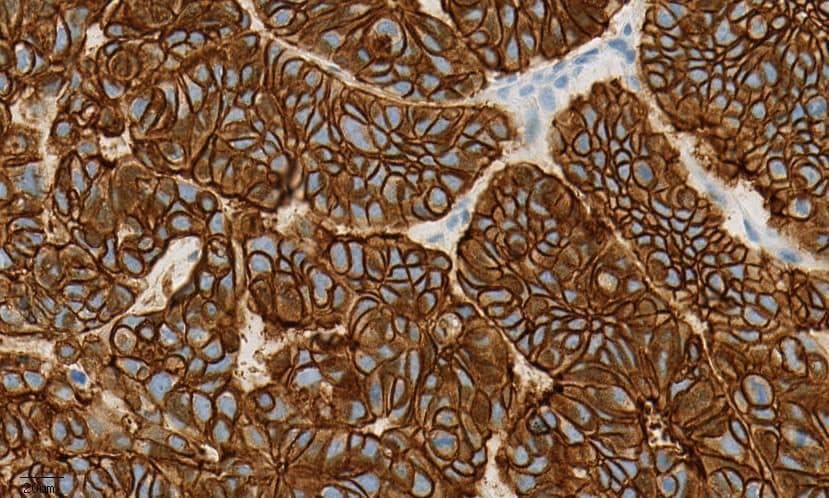
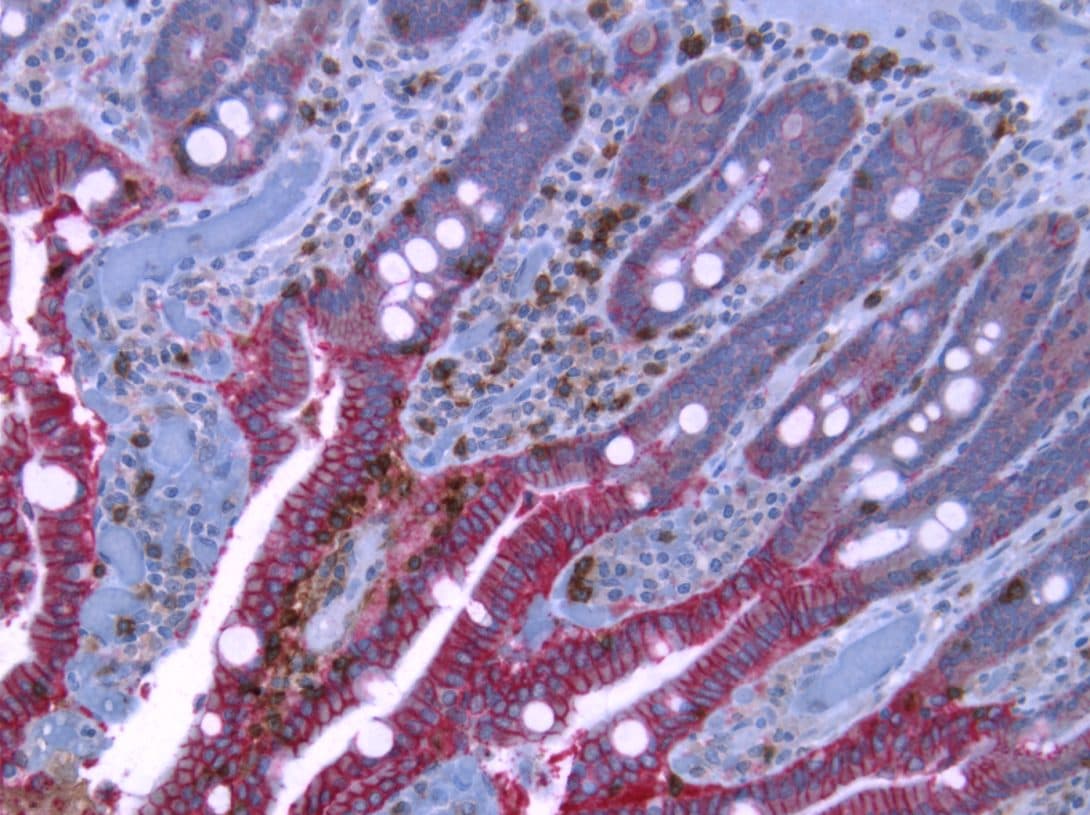
  • Immunohistochemistry

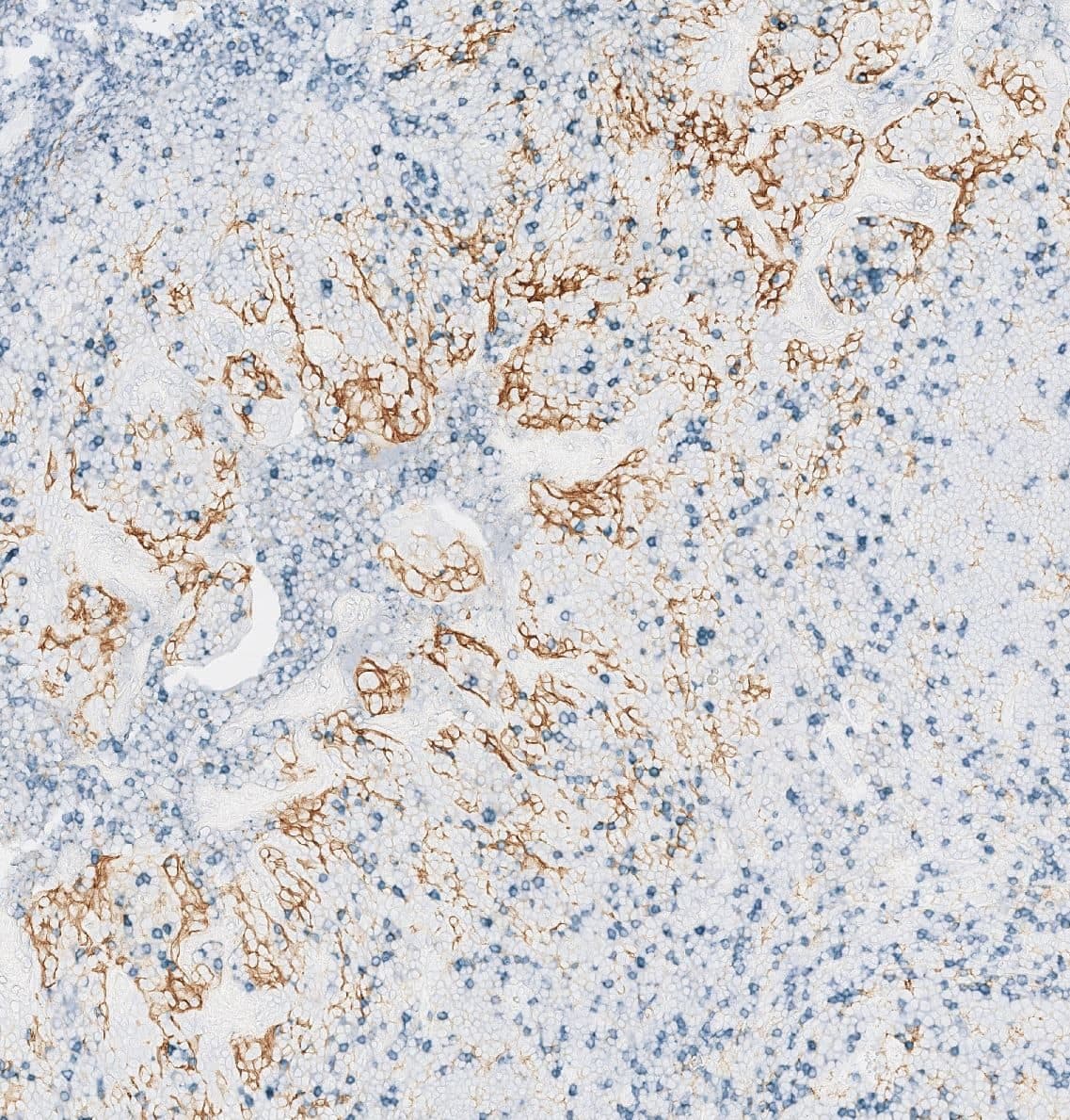
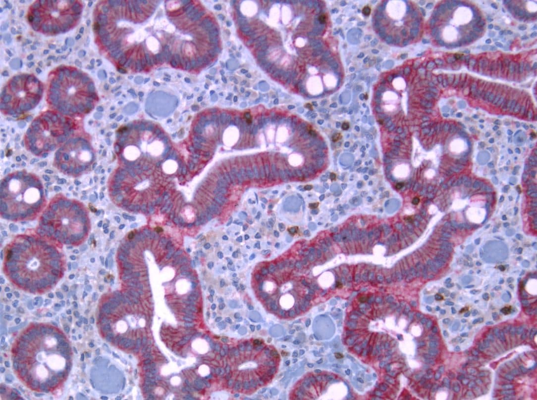
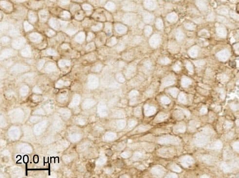
    Single-plex chromogenic IHC across organ systems: routine panels, complex panels, and method development for new targets. Antibody optimization and protocol development for targets without an established protocol.

  • Histology

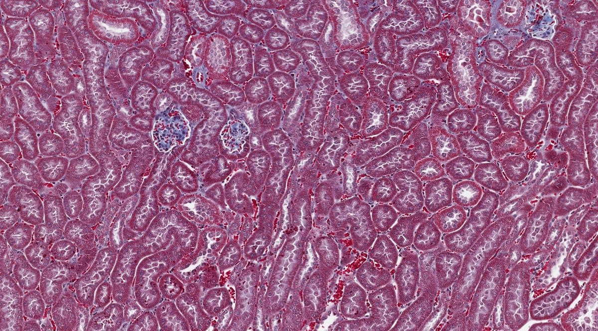
    Accessioning, grossing and trimming, tissue processing, embedding, and sectioning. Standard sections at 4–5 µm on positively charged slides; custom thickness on request. H&E and an extensive special stain menu, with custom stain development available.

  • Pathologist Evaluation

    Qualified pathologist assessment of research specimens, reported as research findings. Available as a standalone service or alongside any staining workflow.

  • Tissue Bank

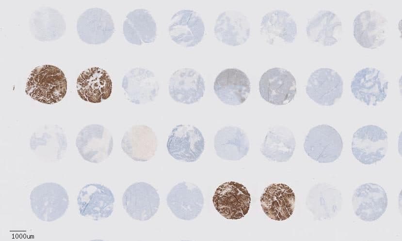
    Research-consented human and animal specimens for preclinical and translational programs, searchable by tissue type and specimen characteristics. Tissue microarray construction available.

Featured services

Preclinical and translational programs, with shared operational excellence.

PathXdx technologists collaborating at laboratory instrumentation

Featured services

Digital pathology & image analysis

Whole-slide scanning and quantitative image analysis in-house: brightfield and fluorescence, digital slide management, and quantitative readouts for marker positivity, co-expression, and spatial relationships.

Common services

Research pathology capabilities for preclinical, discovery, and translational programs: whole-slide imaging and quantitative image analysis, multiplex immunofluorescence, immunohistochemistry, histology (accessioning through H&E and special stains), pathologist evaluation of research specimens, tissue banking, and tissue microarray (TMA) construction.

Our goal

Our goal is to work with you so you meet your goals. We take the extra step to make sure you're happy with the speed and quality of the service we provide.

Our mission

Our mission is advancing discovery. By understanding your study needs we can provide the best solution so you may maximize your potential in the pursuit of your research goals.

How we can help you

When you reach out, we assign a technologist to help design your study, explain the steps, and prepare a proposal. With a PO and timeline, we complete the work and invoice your accounts payable when services are returned.

Discuss your study with PathXDx

Tell us what you're working on. Tissue type, targets, study stage, and timeline. We'll tell you what's feasible and what it costs.