Research use only
Research pathology services
Histology, IHC, multiplex immunofluorescence, pathologist evaluation, and whole-slide scanning for preclinical, discovery, and translational programs. Share your study design and we'll tell you what's feasible, what it costs, and how long it takes before you commit.
Capabilities
What we do
Multiplex immunofluorescence
3–4 plex panels developed and optimized for your targets and tissue, delivered as one workflow rather than handed between vendors.
- Panel design and antibody optimization
- Staining and whole-slide fluorescence imaging
- Panels for immune and tumor microenvironment markers
- Several markers on one section when single-marker IHC isn't enough
Immunohistochemistry
Single-plex chromogenic IHC across organ systems.
- Routine and complex panels
- Method development for new targets
- Antibody optimization and protocol development
Histology
Accessioning through sectioning and staining, for FFPE and frozen workflows.
- Grossing, trimming, processing, and embedding
- 4–5 µm sections on charged slides; custom thickness, serial, and step sections
- H&E and special stains, with custom stain development
- Tissue microarray construction
Pathologist evaluation
Qualified pathologist assessment of research specimens, reported as research findings.
- Standalone, or alongside any staining workflow
- Research-use findings, not diagnostic interpretation
Whole-slide scanning
Brightfield and fluorescence scanning on our own equipment, with whole-slide images (WSI) delivered to you for review, sharing, and your own analysis.
- Brightfield and fluorescence whole-slide scanning
- Whole-slide image files delivered to you
- Digital slide archiving for your program
- Browse tissue blocks
Tissue blocks
Human FFPE tissue by indication, cell pellet blocks, and mouse tissue, plus blocks made from your own cells or animals.
From our lab
Our work
Brightfield IHC, multiplex immunofluorescence, special stains, and whole-slide scans from the PathXDx lab.
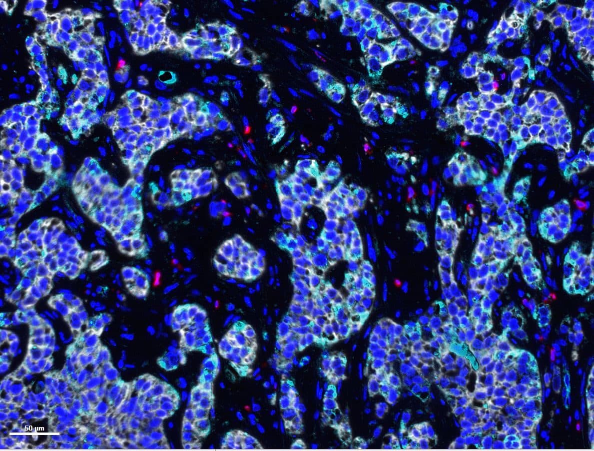
Multiplex immunofluorescence
Tumor
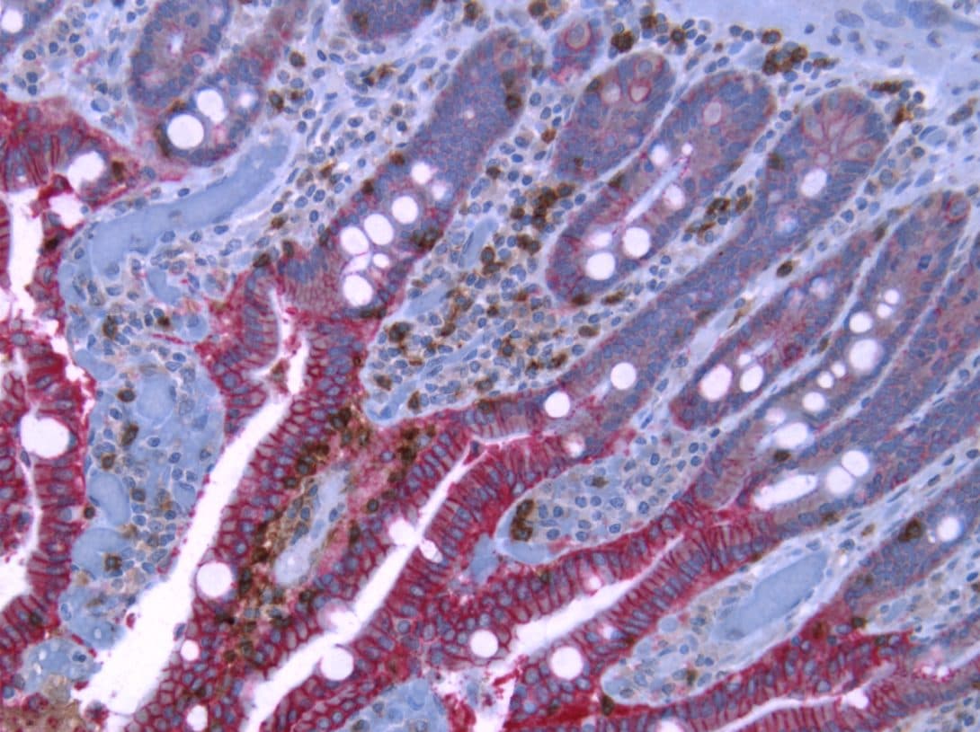
Dual IHC
Human colon cancer
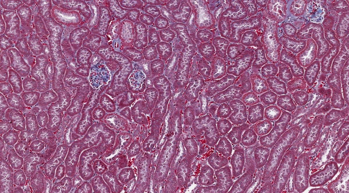
Special stain
Human kidney
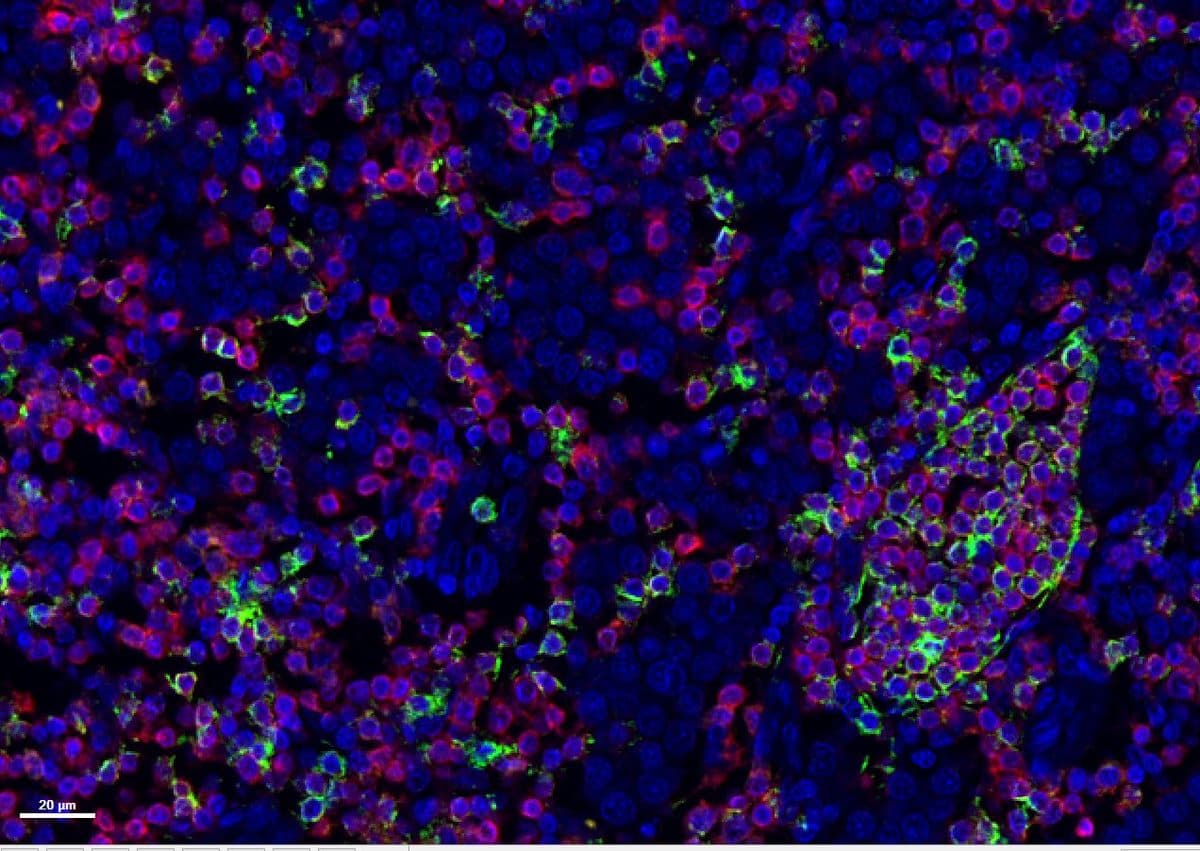
Multiplex immunofluorescence
Lymphoid tissue
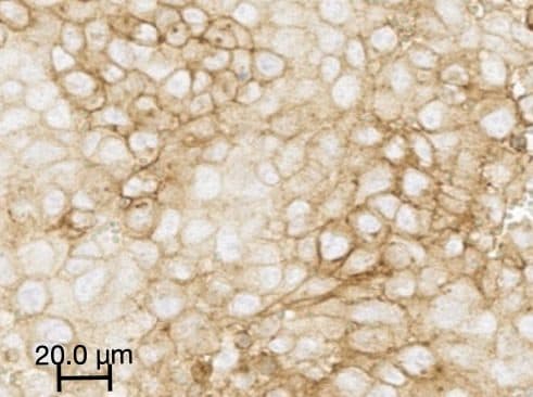
Brightfield IHC
Tumor
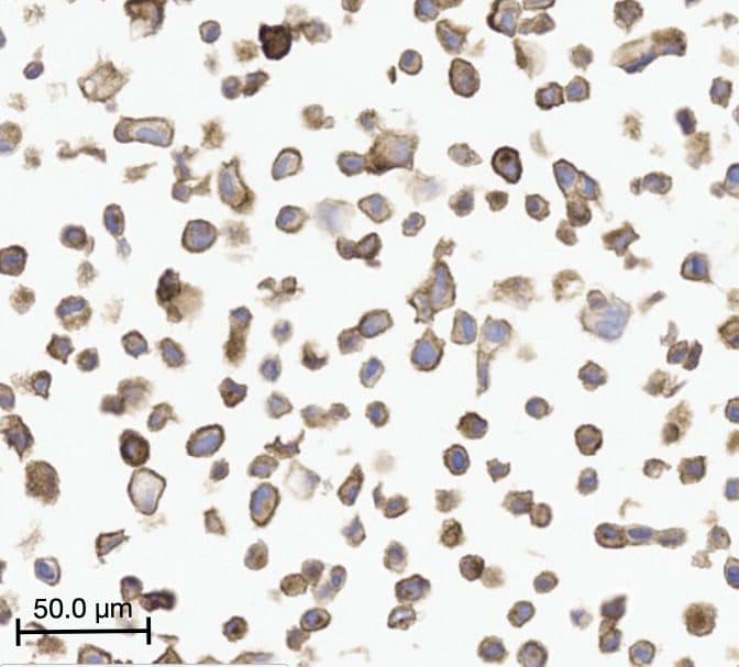
IHC
Cell pellet block
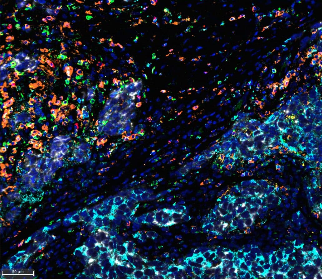
Multiplex immunofluorescence
Tumor microenvironment
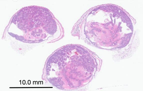
Whole-slide H&E scan
Tissue sections
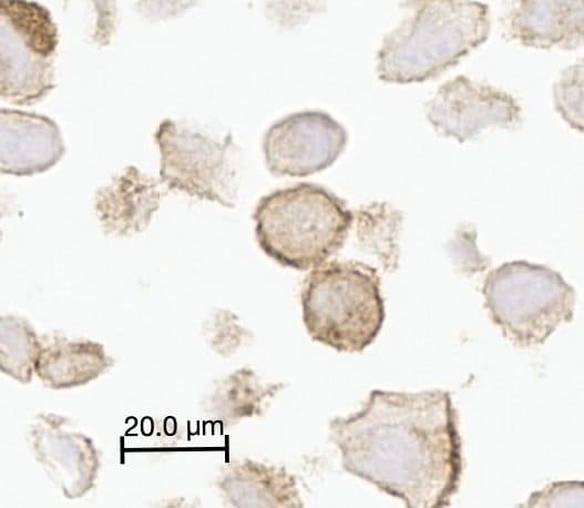
IHC
Cell pellet block, high magnification
Workflow
From receipt to results
How specimens move through the lab, with every step recorded in our LIMS.
- 1
Accessioning & intake
Specimens logged in our LIMS, labeled, and QC'd on receipt, with every step recorded from there.
- 2
Grossing, trimming & processing
Dictation and trimming to your protocol; tissue processing for FFPE or frozen workflows.
- 3
Embedding & sectioning
Standard 4–5 µm sections on positively charged slides. Custom thickness, serial sections, and step sections on request.
- 4
Staining
H&E, special stains, single-plex IHC, or multiplex immunofluorescence, depending on your study.
- 5
Whole-slide scanning
Brightfield and fluorescence scanning on our own equipment. Whole-slide images are delivered to you and archived for your program.
- 6
Pathologist evaluation
Qualified pathologist assessment of research specimens where your study calls for it. Reported as research findings.
- 7
Reporting
Results delivered with methods documented, so your team can interpret and reproduce them.
Sample tracking
Every sample tracked in our LIMS
When you send us a study, each sample and slide is logged in our own laboratory information management system, from receipt to delivery.
Accessioned on arrival
Each sample gets a unique ID, logged with species, tissue, and the services you ordered.
Labeled samples and slides
Every sample and slide carries a printed label that matches its LIMS record.
Every step recorded
Processing, sectioning, and staining steps are logged with who did them and when.
Sample record
Sample 03 · Mouse liver
- Received and accessionedlogged
- Processed and embeddedlogged
- Sectionedlogged
- Stained
- Scanned
Expertise
Organ systems we support
Most tissue types are within scope. Working in an area not listed? Ask us.
- Cardiology
- Dermatology
- Gastroenterology
- Gynecology
- Hematology
- Musculoskeletal
- Nephrology
- Neurology
- Ophthalmology
- Otolaryngology
- Urology
- Special projects
Why PathXDx
High-touch partnership, owned imaging capability
Everything under one roof
Histology, staining, and whole-slide scanning happen in our own lab. No pass-through vendors, no queue behind someone else's work, and one point of accountability from block to slide.
Multiplex without building it yourself
Multiplex immunofluorescence takes real optimization to get right. We've done that work, so you don't need to stand up the capability internally for a single study.
A technologist who knows your protocol
Named technologists are assigned to your program. They learn your tissue, your protocol, and your acceptance criteria.
Straight answers on scope
If a study design won't produce the data you need, we say so before you commit, not after.
Ready to scope your study?
Tell us the tissue type, targets, study stage, and timeline. A technologist will help design the study and prepare a proposal.
Discuss your study